
Located between the lungs, your heart is in charge of pumping blood within the body through the blood vessels. This blood will provide nutrients and oxygen for your body, as well as carrying wastes from metabolism processes.
The right part of your heart pumps blood to the lungs: receiving oxygen. Meanwhile, the left one receives oxygen-induced blood and pumps it to the rest of your body.
In humans, birds, as well as mammals, the heart is divided into four chambers or compartments: the right ventricle, the left atrium, the left ventricle, and the right atrium. Divided into four valves—one of each compartment of the heart, the heart valves ensure that your blood moves in the correct direction. The opening and closing of each valve depend on the different pressure in the compartment.
Every part of your heart is vital to your life. Therefore, it is essential to learn how heart valves work. Knowing heart valves functions and anatomy is beneficial in understanding its task in providing a normal and healthy circulation.
Heart Valves Anatomy
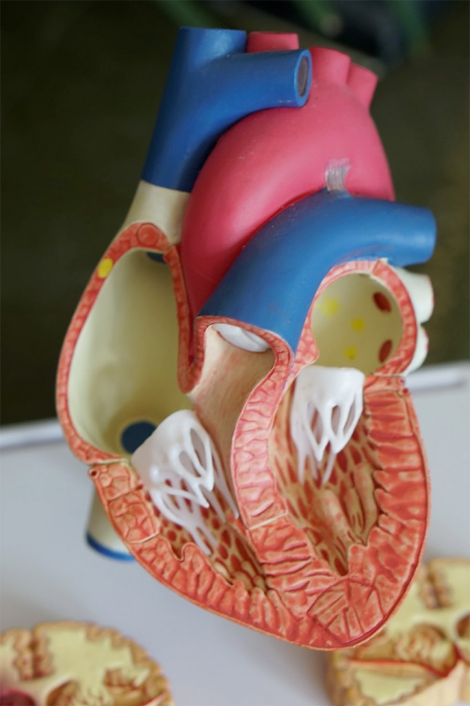
Made of tough, thin tissues named leaflets or cusps, these leaflets or cusps opens during the half heartbeat to let the blood move within. Afterwards, they close during the other half of the heartbeat to ensure your blood from going backwards.
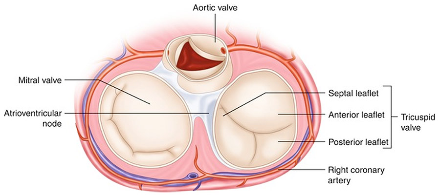
The mitral and tricuspid valves each have two cusps. Meanwhile, aortic and pulmonary have three. These leaflets are attached to the valves by the annulus of the heart, made out of a ring of fibrous tissue.
Human’s heart valves can be grouped into two parts: two Atrioventricular Valves and two Semilunar Valves. Consist of the bicuspid or mitral valve and tricuspid valve, your atrioventricular valves are accountable to halt the blood course going reverse into the atria from the ventricles.
In contrast, semilunar valves are in charge of halting your blood from going reverse to the ventricle. These valves are divided into two parts, which are pulmonary valve and aortic valve.
How Heart Valves Work
The four heart valves are working hand-in-hand to ensure that your blood is running in the correct direction as well as to stop blood leakage in your ventricle or artery. Here are the steps of how blood circulation in your heart works.
Mitral and Tricuspid Valves are open
Through the opened tricuspid valve, the blood is flowing from the right atrium into the right ventricle. Afterward, to the left ventricle from the left atrium through the opened mitral valve.
Mitral and Tricuspid Valves are closed
Once the blood in the ventricle is full, the mitral and tricuspid valves close to halt your blood course going reverse thus creating leakage during contractions. The tricuspid valve is responsible for the right ventricle while the mitral valve for the left ventricle.
Aortic and Pulmonary Valves are open
During contraction within the ventricle, your aortic and pulmonary valves compel to open by the blood course. Your pulmonary valve pumps blood in the right ventricle to the pulmonary artery.
In contrast, blood in the left ventricle is pumped to the aorta through your aortic valve. Aorta itself consists of numerous arteries that will take the blood throughout your body.
Aortic and Pulmonary Valves are closed
Once finished contracting, both ventricles will begin relaxing and your aortic and pulmonary valve shutting off. This closure halts blood from running backwards, which can result in an unwanted leakage in your heart.
Although it seems extensive, these movements are repeated continuously in a short period of the heartbeat. The continuous movements ensure that blood will course to your heart, body, and lungs without problems. Normal heart valves direct the blood in the correct way as well as shut at the exact time to stop leakage.
Heart Valves Functions
Here are human heart valves functions in accordance with the circulation course:
Tricuspid Valve
Situated on the right part of the atrioventricular valve, your tricuspid valve functions to halt blood backflow during contractions from the right ventricle to the right atrium. The tricuspid valve is made up of three leaflets, an annulus, and supportive features which are papillary muscles and chordae tendineae.
Also known as heart-string, chordae tendineae is a strong, fibrous strand that attaches papillary muscles to the mitral and tricuspid valve. Meanwhile, papillary muscle is a muscle located within the walls of your heart’s ventricles.
Two most familiar congenital tricuspid valve diseases are tricuspid atresia and Ebstein’s anomaly. Someone who suffers from tricuspid atresia doesn’t have a tricuspid valve, resulting in a solid obstruction between your right heart compartments.
Additionally, Ebstein’s anomaly means that you have a malformed tricuspid valve, in which your valve sits lower than usual in the right ventricle, causing a backflow to the correct atrium.
Aside from tricuspid atresia and Ebstein’s anomaly, someone can suffer from tricuspid valve stenosis, in which your valve is too cramped that the blood course from the right atrium to the lower right heart compartment is obstructed.
Pulmonary Valve
Separating the right ventricle from the pulmonary artery, your pulmonary valve is semilunar consisting of three cusps. Located in anterior, superior, and close to the left of the aortic valve, pulmonary valve’s role is to stop blood from retracting reverse to the right ventricle from the pulmonary artery.
A condition which disturbs pulmonary valve’s performance is called pulmonary stenosis, in which your blood is obstructed in the pulmonary valve. Congenital heart disease is a common cause of this problem, along with rheumatic heart disease and malignant carcinoid tumour.
The best way to treat pulmonary stenosis is through surgery or replacement of the valve.
Mitral Valve
Also known as the bicuspid valve or left atrioventricular valve, your mitral valve is accountable in delivering blood from the left atrium to the left ventricle during contractions. Comprises of two cusps, mitral valve and tricuspid valve are collectively known as Atrioventricular Valves (AV) due to its position, between your heart’s ventricles and atria.
Similar to the tricuspid valve, papillary muscles and chordae tendineae also support your mitral valve. Additionally, the mitral valve also has an annulus to ensure that the blood won’t run reverse to the left atrium during contractions.
The mitral valve also has several conditions that need to be noted. Mitral stenosis occurs when your mitral valve is cramped, creating a blockage of the blood course. Causing a shortage of breath and tiredness, mitral stenosis is usually caused by rheumatic fever, which scars the mitral valve.
Additionally, another mitral valve condition is mitral valve prolapse. Mitral valve prolapse occurs when your mitral valve slips reverse into the left atrium of the heart, causing a small amount of blood leakage to the atrium. This is caused by either size abnormalcy or damage on the mitral valve.
Although harmless, a serious mitral valve prolapse can cause an abnormal heartbeat which leads to a chronic issue.
Aortic Valve
Also called the semilunar valves of the heart, aortic valve separates the aorta and heart’s left ventricle. Comprises of two or three cusps, the aortic valve is the last stop of heart’s blood circulation before the blood is delivered through numerous arteries in your aorta.
Aortic valve disease is among the most familiar heart valves diseases, in which the left ventricle of the heart and the aorta doesn’t work correctly. The cause ranges from congenital or birth-conditioned or other varied causes. Two most likely diseases are aortic valve stenosis and aortic valve regurgitation.
Often caused by rheumatic fever, aortic valve stenosis is a condition where your aortic valve becomes tough and stiff, thus blocking the blood course from the aorta to your body. Whereas, aortic valve regurgitation occurs when the aortic valve isn’t closed appropriately, causing a blood leakage to your heart’s left ventricle.
Aortic valve diseases don’t usually show apparent symptoms for years. It usually shows in the form of shortage of breath, tiredness, fainting, and irregular heartbeat, among many others.
You can opt to either repair or replace the aortic valve once diagnosed. Disease such as aortic valve regurgitation could be treated through reconstruction and treatment.
In contrast, aortic valve replacement replaces a native valve with a prosthetic one, made of either metal or animal tissues. The replacement occurs through the surgical or non-surgical method.
Heart valves are one of many vital organs in the human’s body. Not only that it is responsible for keeping your body alive, but fully functioning heart valves also regulate the quality of your health.


Leave a Reply